
More Information
Submitted: 13 October 2020 | Approved: 30 October 2020 | Published: 02 November 2020
How to cite this article: Briceño AB, Oliva H. Gastrointestinal stromal tumor resulting in recurrent colic in a arabian horse gelding a report of case. Insights Vet Sci. 2020; 4: 048-050.
DOI: 10.29328/journal.ivs.1001026
ORCiD ID: orcid.org/0000-0002-9122-9251
Copyright License: © 2020 Briceño AB, et al. This is an open access article distributed under the Creative Commons Attribution License, which permits unrestricted use, distribution, and reproduction in any medium, provided the original work is properly cited.
Keywords: Arabian; Colic; Horse; Recurrent; Stromal

Gastrointestinal stromal tumor resulting in recurrent colic in a arabian horse gelding a report of case
Abelardo Morales Briceño* and Harmon Oliva
SHS Veterinary Center, Al Wathba, Abu Dhabi, United Arab Emirates
*Address for Correspondence: Abelardo Morales Briceño, SHS Veterinary Center, Al Wathba, Abu Dhabi, United Arab Emirates, Tel: +34619307223; Email: [email protected]
Background: A Grey 12-year-old Arabian endurance horse gelding was referred to the SHS Veterinary Center for anorexia, mild colic of 5 days duration, and melena of 1 day duration. The owner reported recurring colic, 12 episodes of mild colic in the previous year.
Methods: On admission, vital signs were within normal limits and body condition score was estimated to be 3/9.
Results: Packed cell volume (PCV) was 28% [reference range (RR): 31% to 47%] and plasma total protein was 58 g/L (RR: 60 to 80 g/L). Hematochezia was observed. Abdominal ultrasound examination detected no abnormalities. Over the next 12 h, the horse experienced hematochezia and several mild episodes of colic and death. A necropsy was performed. A mass arising from the right dorsal ascending colon near the base of the cecum and extending transmurally from the colonic mucosa into the mesocolon was a 8 cm × 5 cm × 8 cm firm, homogenous, tan mass. The portion of the mass that extended into the colonic lumen was pedunculated, with an ulcerated surface. The adjacent segments of colon were markedly reddened and edematous. Histologically, the mass was comprised of large interweaving sheets of small, spindle cells with ill-defined cell borders embedded in abundant myxomatous matrix. Tumor cells contained scant eosinophilic cytoplasm and oval to elongate nuclei with finely stippled chromatin and inconspicuous nucleoli. Mitotic figures were rare (1/10) high power fields. Tumor infiltrated between the muscularis interna and the muscularis externa at the myenteric plexi.
Conclusion: Gross and histologic appearance, were consistent with a diagnosis of gastrointestinal stromal tumor.
A recently study identified and described the causes of death in Arabian horses, in summary, pathologies of the digestive system, specifically acute abdominal crisis (colic), are the main causes of death in Arabian horses (80%) [1]. The gastro-intestinal tract and abdominal organs are susceptible to a variety of primary tumours and metastatic tumours [2]. Gastrointestinal tumors in horses are rare; however, in some cases paraneoplastic signs such as recurrent colic, protein loss syndrome, and emaciation, among others associated with the presence of tumors, may occur. The most common neoplasias of the equine intestinal tract are lymphoma, adenocarcinoma, and leiomyosarcoma and Gastrointestinal Stromal Tumors (GIST) are rare in horses and few reports exist [3]. Additionally, lipomas and liposarcomas in horses are incidental findings at necropsy and are not associated with preceding clinical signs in some cases, but they represent a risk factor for the development of acute abdominal crisis and the effect of mass occupying space in the abdominal cavity. A case of severed infiltrative lipomatosis intestinal in large colon and in dorsal neck muscle with deformation of dorsal neck and colic in a Spanish Pure Breed Horse was report [4]. Is true to say that intestine or neoplasia is rare but early diagnosis is critical if any success is to be achieved in respect of treatment [2]. The aim of this study was to report a Gastrointestinal Stromal Tumor (GIST) resulting in recurrent colic in a Arabian horse Gelding.
A Grey 12-year-old Arabian endurance horse gelding was referred to the SHS Veterinary Center for anorexia, mild colic of 5 days duration, and melena of 1 day duration. The owner reported recurring colic, 12 episodes of mild colic in the previous year. On admission, vital signs were within normal limits and body condition score was estimated to be 3/9. Packed cell volume (PCV) was 28% [reference range (RR): 31% to 47%] and plasma total protein was 58 g/L (RR: 60 to 80 g/L). Hematochezia was observed. Abdominal ultrasound examination detected no abnormalities. Over the next 12h, the horse experienced hematochezia and several mild episodes of colic and death. A necropsy was performed, a mass arising from the right dorsal ascending colon near the base of the cecum and extending transmurally from the colonic mucosa into the mesocolon was 8 cm × 5 cm × 8 cm firm, homogenous (Figure 1). The portion of the mass that extended into the colonic lumen was pedunculated, with an ulcerated surface (Figures 2,3). The adjacent segments of colon were markedly reddened and edematous. Histologically, the mass was comprised of large interweaving sheets of small, spindle cells with ill-defined cell borders embedded in abundant myxomatous matrix. Tumor cells contained scant eosinophilic cytoplasm and oval to elongate nuclei with finely stippled chromatin and inconspicuous nucleoli. Mitotic figures were rare (1/10) high power fields. Tumor infiltrated between the muscularis interna and the muscularis externa at the myenteric plexi. Gross and histologic appearance, were consistent with a diagnosis of gastrointestinal stromal tumor.
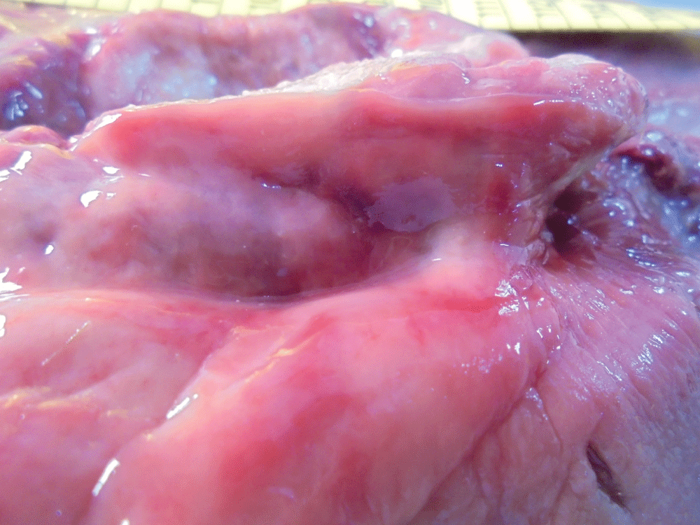
Figure 1: A mass arising from the right dorsal ascending colon near the base of the cecum and extending transmurally from the colonic mucosa into the mesocolon was 8 cm × 5 cm × 8 cm firm, homogenous.JPG
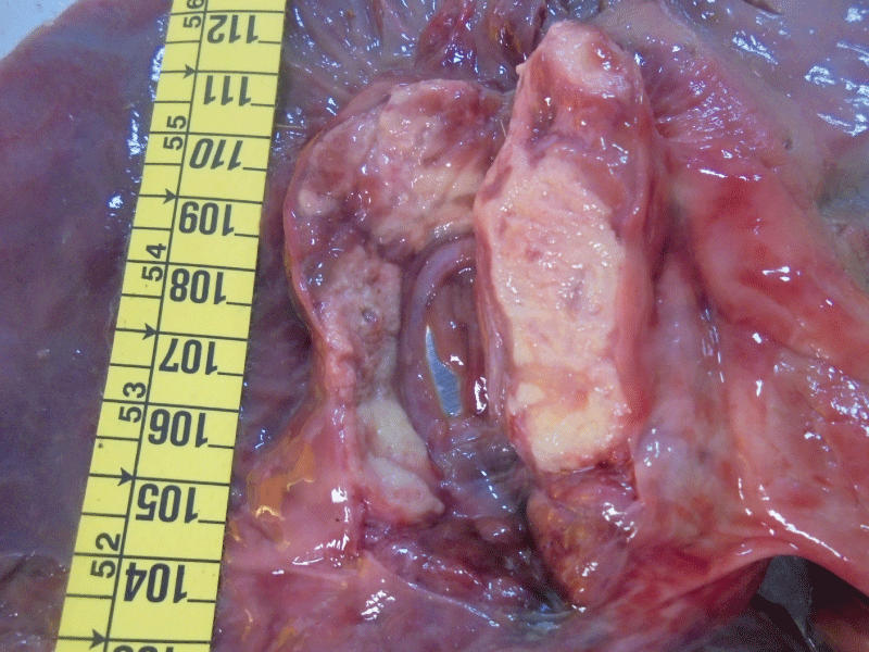
Figure 2: The portion of the mass that extended into the colonic lumen was pedunculated, with an ulcerated surface.JPG
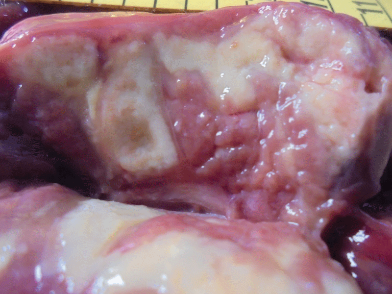
Figure 3: The portion of the mass that extended into the colonic lumen was pedunculated, with an ulcerated surface.JPG
GISTs are rather uncommon tumors in horses. This type of tumor can manifest clinically with paraneoplastic signs such as recurrent colic, as well as anemia and melena. Abdominal neoplasia has been implicated in around 4% of horses presenting with intermittent or chronic colic signs [2]. In other cases, GISTs may be an incidental necropsy finding. Most of these tumors are comprised of a loosely arranged network of spindled cells separated by myxoid matrix, GIST may be composed of myogenic, neurogenic, combined myogenic and neurogenic, and undifferentiated mesenchymal cells [5]. Gastrointestinal stromal tumors (GISTs) are defined as specific CD117-(Kit, stem cell factor receptor) expressing tumors of the gastrointestinal (GI) tract, they are believed to originate from the interstitial pacemaker cells of Cajal (ICC) or their progenitor cells [6]. The diagnosis in this case was carried out a clinical, macroscopic and histological study of the GIST. Ten cecal tumors were identified during the postmortem examination of seven horse carcasses at slaughter (one horse had three tumors) [7]. A study of 10 gastrointestinal stromal tumors in equids in Thoroughbred and Appaloosa breeds and 1 pony, examined grossly, histologically, immunohistochemically, and (in two cases) ultrastructurall [5], a 24-year-old, 540 kg Warm-Blood gelding [3], and a 14-year-old Trakehner gelding [6]. Although there is marked tendency for neoplastic disease to be increasingly prevalent in older aged horses, gastrointestinal tumours are not the preserve of older animals [2]. This report describes the first case of an Arabian horse Gastrointestinal Stromal Tumor (GIST). In many cases, the diagnostic imaging can be useful to identify a tumor mass and take a sample for cytological study and biopsy, the surgical option may represent a therapeutic alternative. Every now and again however there is one that causes a serious intestine obstruction - usually of a strangulating nature requiring emergency intervention, removal of focal haemangiosarcoma, leiomyoma, and even focal intestinal lymphoma tumours can be undertaken with success [2]. The equine cecal stromal tumors described in this report represent a distinctive group of mesenchymal tumors [7]. Microscopic examination revealed discrete spindle cells arranged in compact patterns with fascicles and whorls or cribriform pattern with fascicles and rare palisades, often with a myxoid interstitial matrix [5]. In conclusion gross and histologic appearance, were consistent with a diagnosis of gastrointestinal stromal tumor.
- Morales-Briceño A. A Retrospective Study of Mortality Causes in Arabian Horses. Rev Med Vet. 2020; 41: 1-15.
- Knottenbelt D, Leverhulme P. Gastrointestinal neoplasia. XX SIVE International Congress, Milano. 2014; 152-160.
- Malberg JA, Webb BT, Hackett E. Colonic gastrointestinal stromal tumor resulting in recurrent colic and hematochezia in a warmblood gelding. Can Vet J. 2014; 55: 471–474. PubMed: https://pubmed.ncbi.nlm.nih.gov/24790234/
- Morales A. Mendez A, Perez Arevalo J. Infiltrative lipomatosis in the neck and colon of a Spanish Pure Breed Horse. Braz J Vet Pathol. 2015; 8: 170-173.
- Del Piero F, Summers BA, Cummings JF, Mandelli G, Blomme EA. Gastrointestinal Stromal Tumors in Equids. Vet Pathol. 2001; 38: 689-697. PubMed: https://pubmed.ncbi.nlm.nih.gov/11732803/
- Stephan S, Hug S, Hilbe M. Gastrointestinal Stromal Tumor in the Cecum of a Horse. Case Reports in Veterinary Medicine. 2012; 301498.
- Hafner S, Harmon BG, King T. Gastrointestinal stromal tumors of the equine cecum. Vet Pathol. 2001; 38: 242-246. PubMed: https://pubmed.ncbi.nlm.nih.gov/11280386/