Figure 4
Comparative anatomy of selected bones of forelimb of local Mongrelian Dog (Canis lupus familiaris) in Sokoto, Nigeria
Bello A* and Wamakko HH
Published: 14 December, 2021 | Volume 5 - Issue 1 | Pages: 026-031
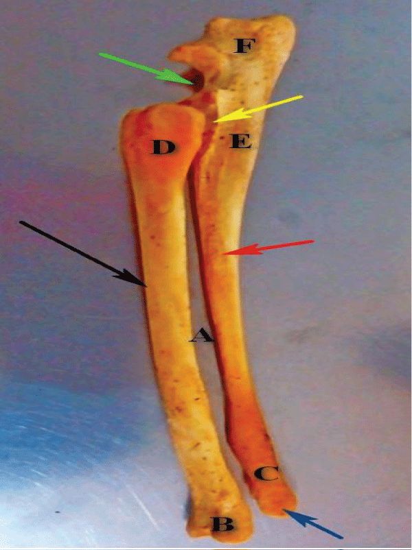
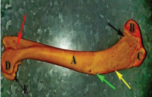
Figure 4:
Photograph of the lateral view of radio ulnar bones of Mongrelian Dog at 2-3 years showing the shaft of the radial bone (black arrow), shaft of the ulna bone (red arrow), proximal extremity of the radius (D), proximal extremity of the ulna (E), the large interoceous space (A), the distal extremity of the radius (B), the distal extremity of the ulna bone (C), the ancornial process (green arrow) and the distal styloid process of the ulna bone (blue arrow).
Read Full Article HTML DOI: 10.29328/journal.ivs.1001033 Cite this Article Read Full Article PDF
More Images
Similar Articles
-
Anatomical changes of the development of red Sokoto goat stomachBello A*,Joseph OA,Onua JE,Onyeanusib BI,Umaru MA,Bodinga HA. Anatomical changes of the development of red Sokoto goat stomach. . 2020 doi: 10.29328/journal.ivs.1001019; 4: 004-009
-
Comparative anatomy of selected bones of forelimb of local Mongrelian Dog (Canis lupus familiaris) in Sokoto, NigeriaBello A*,Wamakko HH. Comparative anatomy of selected bones of forelimb of local Mongrelian Dog (Canis lupus familiaris) in Sokoto, Nigeria. . 2021 doi: 10.29328/journal.ivs.1001033; 5: 026-031
Recently Viewed
-
Comparative characterization between autologous serum and platelet lysate under different temperatures and storage timesCamilo Osorio Florez*, Luis Campos, Jessica Guerra, Henrique Carneiro, Leandro Abreu, Andres Ortega, Fabiola Paes, Priscila Fantini, Renata de Pino Albuquerque Maranhão. Comparative characterization between autologous serum and platelet lysate under different temperatures and storage times. Insights Vet Sci. 2023: doi: 10.29328/journal.ivs.1001038; 7: 001-009
-
A Perspective on Conservation Technologies for Endangered Marine BirdsAnn Morrison*, Sonja Lukaszewicz. A Perspective on Conservation Technologies for Endangered Marine Birds. Insights Vet Sci. 2023: doi: 10.29328/journal.ivs.1001039; 7: 010-014
-
Intrauterine Therapy with Platelet-Rich Plasma for Persistent Breeding-Induced Endometritis in Mares: A ReviewThiago Magalhães Resende*,Renata Albuquerque de Pino Maranhão,Ana Luisa Soares de Miranda,Lorenzo GTM Segabinazzi,Priscila Fantini. Intrauterine Therapy with Platelet-Rich Plasma for Persistent Breeding-Induced Endometritis in Mares: A Review. Insights Vet Sci. 2024: doi: 10.29328/journal.ivs.1001045; 8: 039-047
-
Comparing Immunity Elicited by Feedback and Titered Viral Inoculation against PEDV in SwineMaría Elena Trujillo Ortega,Selene Fernández Hernández,Montserrat Elemi García Hernández,Rolando Beltrán Figueroa,Francisco Martínez Castañeda,Claudia Itzel Vergara Zermeño,Sofía Lizeth Alcaráz Estrada,Elein Hernández Trujillo,Rosa Elena Sarmiento Silva*. Comparing Immunity Elicited by Feedback and Titered Viral Inoculation against PEDV in Swine. Insights Vet Sci. 2024: doi: 10.29328/journal.ivs.1001044; 8: 028-038
-
Comparative Analysis of Water Wells and Tap Water: Case Study from Lebanon, Baalbeck RegionChaden Moussa Haidar, Ali Awad, Walaa Diab, Farah Kanj, Hassan Younes, Ali Yaacoub, Marwa Rammal, Alaa Hamze. Comparative Analysis of Water Wells and Tap Water: Case Study from Lebanon, Baalbeck Region. Insights Vet Sci. 2024: doi: 10.29328/journal.ivs.1001043; 8: 018-027
Most Viewed
-
Impact of Latex Sensitization on Asthma and Rhinitis Progression: A Study at Abidjan-Cocody University Hospital - Côte d’Ivoire (Progression of Asthma and Rhinitis related to Latex Sensitization)Dasse Sery Romuald*, KL Siransy, N Koffi, RO Yeboah, EK Nguessan, HA Adou, VP Goran-Kouacou, AU Assi, JY Seri, S Moussa, D Oura, CL Memel, H Koya, E Atoukoula. Impact of Latex Sensitization on Asthma and Rhinitis Progression: A Study at Abidjan-Cocody University Hospital - Côte d’Ivoire (Progression of Asthma and Rhinitis related to Latex Sensitization). Arch Asthma Allergy Immunol. 2024 doi: 10.29328/journal.aaai.1001035; 8: 007-012
-
Causal Link between Human Blood Metabolites and Asthma: An Investigation Using Mendelian RandomizationYong-Qing Zhu, Xiao-Yan Meng, Jing-Hua Yang*. Causal Link between Human Blood Metabolites and Asthma: An Investigation Using Mendelian Randomization. Arch Asthma Allergy Immunol. 2023 doi: 10.29328/journal.aaai.1001032; 7: 012-022
-
An algorithm to safely manage oral food challenge in an office-based setting for children with multiple food allergiesNathalie Cottel,Aïcha Dieme,Véronique Orcel,Yannick Chantran,Mélisande Bourgoin-Heck,Jocelyne Just. An algorithm to safely manage oral food challenge in an office-based setting for children with multiple food allergies. Arch Asthma Allergy Immunol. 2021 doi: 10.29328/journal.aaai.1001027; 5: 030-037
-
Snow white: an allergic girl?Oreste Vittore Brenna*. Snow white: an allergic girl?. Arch Asthma Allergy Immunol. 2022 doi: 10.29328/journal.aaai.1001029; 6: 001-002
-
Cytokine intoxication as a model of cell apoptosis and predict of schizophrenia - like affective disordersElena Viktorovna Drozdova*. Cytokine intoxication as a model of cell apoptosis and predict of schizophrenia - like affective disorders. Arch Asthma Allergy Immunol. 2021 doi: 10.29328/journal.aaai.1001028; 5: 038-040

If you are already a member of our network and need to keep track of any developments regarding a question you have already submitted, click "take me to my Query."


