More Info
Author's satisfaction with
- Friendly and hassle-free publication process
- Less production time of articles
- Constructive peer-review
- Enhancing journal reputation
- Regular feedback system
- Quick response to authors' queries
Recently Viewed
Most Viewed
Clinical Images
Abstract
Research Article
Comparative characterization between autologous serum and platelet lysate under different temperatures and storage times
Camilo Osorio Florez*, Luis Campos, Jessica Guerra, Henrique Carneiro, Leandro Abreu, Andres Ortega, Fabiola Paes, Priscila Fantini and Renata de Pino Albuquerque Maranhão
Published: 12 April, 2023 | Volume 7 - Issue 1 | Pages: 001-009
Therapies using autologous serum and platelet lysate have shown promise among blood and biological products in the treatment of various diseases. The autologous serum has been shown to be a superior alternative to traditional eye drops in treating eye diseases in ophthalmology. Platelet lysate (PL) has recently been considered a more interesting alternative for the treatment of multiple tissues, as it does not have the unfavorable reactions seen with traditional platelet-rich plasma (PRP), making it a valuable blood derivative for use in ocular therapy. There is no definitive comparison in veterinary medicine between PL and autologous serum in terms of the content of Transforming Growth Factor beta 1 (TGF-1), which is known to have chemotactic, mitogenic, matrix formation, and angiogenesis effects on tissues, and beneficial proteins in ocular tissue. This study aimed to estimate the concentrations of TGF-1, total protein, and albumin, as well as autologous serum and platelet lysate, in horses over an 8-day storage period at temperatures of 4 °C and 37 °C.
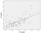
To produce autologous serum, 63 ml of blood was collected from each animal in seven 9 ml tubes without anticoagulant. For platelet lysate, 180 ml of blood was collected in 50 tubes of 3.6 ml with 3.2% sodium citrate. The most significant findings were the positive relationship between the baseline platelet count in the blood and the final platelet concentration in PRP. Specifically, we found a correlation (R = 0.9) with a p - value of 0.005 between the average baseline platelet level of seven animals and their corresponding PRP results, both on an individual level and as a group. Additionally, there was a correlation between growth factor concentration and PRP platelets, with the highest growth factor concentration in PL. The temperature storage group exhibited higher concentrations of total protein and serum albumin, as well as the maximum amount of growth factor for both products at a temperature of 37 °C.
Read Full Article HTML DOI: 10.29328/journal.ivs.1001038 Cite this Article Read Full Article PDF
References
- Maia L, De Souza MV. Components rich in platelets used in wound healing tendon, ligaments and osteo-articular diseases of animals. Ciência Rural. 2009; 39: 1279–1286.
- De Souza MV, Pinto O, Da Costa MM. Quantification of growth factors in horse skin treated with platelet-rich plasma. 2014; 34: 599–612.
- Mirza AUB, Ghani N, Khan AB. Epitheliotrophic Effect of Autologous Serum in Persistent Corneal Epithelial Defects. Pakistan Journal of Ophthalmology. 2008; 24:19-26.
- López-García JS, García-Lozano I, Rivas L, Martínez-Garchitorena J. Aplicaciones del suero autólogo en oftalmología [Use of autologous serum in ophthalmic practice]. Arch Soc Esp Oftalmol. 2007 Jan;82(1):9-20. Spanish. doi: 10.4321/s0365-66912007000100004. PMID: 17262232.
- Pan Q, Angelina A, Marrone M, Stark WJ, Akpek EK. Autologous serum eye drops for dry eye. Cochrane Database Syst Rev. 2017 Feb 28;2(2):CD009327. doi: 10.1002/14651858.CD009327.pub3. PMID: 28245347; PMCID: PMC5510593.
- Mandelbaum SH, Di Santi, É, Mandelbaum M. Cicatrization: current concepts and auxiliary resources - Part II. An Bras Dermatol. 2003; 78: 525–542.
- Paganela JC, Ribas LM, Santos CA. Abordagem clínica de feridas cutâneas em equinos Clinical approach in equine skin wounds. Revista Portuguesa de Ciências Veterinárias. 2009; 104: 569–572.
- Anitua E, Muruzabal F, Tayebba A, Riestra A, Perez VL, Merayo-Lloves J, Orive G. Autologous serum and plasma rich in growth factors in ophthalmology: preclinical and clinical studies. Acta Ophthalmol. 2015 Dec;93(8):e605-14. doi: 10.1111/aos.12710. Epub 2015 Apr 2. PMID: 25832910.
- Schallmoser K, Henschler R, Gabriel C, Koh MBC, Burnouf T. Production and Quality Requirements of Human Platelet Lysate: A Position Statement from the Working Party on Cellular Therapies of the International Society of Blood Transfusion. Trends Biotechnol. 2020 Jan;38(1):13-23. doi: 10.1016/j.tibtech.2019.06.002. Epub 2019 Jul 17. PMID: 31326128.
- Soares CS, Babo PS, Reis RL, Carvalho PP, Gomes ME. Platelet-Derived Products in Veterinary Medicine: A New Trend or an Effective Therapy? Trends Biotechnol. 2021 Mar;39(3):225-243. doi: 10.1016/j.tibtech.2020.07.011. Epub 2020 Aug 28. PMID: 32868100.
- Penha-Goncalves MN, Onions DE, Nicolson L. Cloning and sequencing of equine transforming growth factor-beta 1 (TGF beta-1) cDNA. DNA Seq. 1997;7(6):375-8. doi: 10.3109/10425179709034059. PMID: 9524819.
- Argüelles D, Carmona JU, Pastor J, Iborra A, Viñals L, Martínez P, Bach E, Prades M. Evaluation of single and double centrifugation tube methods for concentrating equine platelets. Res Vet Sci. 2006 Oct;81(2):237-45. doi: 10.1016/j.rvsc.2005.12.008. PMID: 16969921.
- Watts EJ, Rose MT. Platelet-derived growth factor acts via both the Rho-kinase and p38 signaling enzymes to stimulate contraction in an in vitro model of equine wound healing. Domest Anim Endocrinol. 2010 May;38(4):253-9. doi: 10.1016/j.domaniend.2009.11.004. Epub 2009 Dec 6. PMID: 20036481.
- Sundman EA, Cole BJ, Fortier LA. Growth factor and catabolic cytokine concentrations are influenced by the cellular composition of platelet-rich plasma. Am J Sports Med. 2011 Oct;39(10):2135-40. doi: 10.1177/0363546511417792. Epub 2011 Aug 16. PMID: 21846925.
- Sanak F, Baenninger P, Kaufmann C, Iselin K, Bachmann L, Buhl D, Thiel M. The Lucerne Protocol for the Production of Autologous Serum Eyedrops. Klin Monbl Augenheilkd. 2021 Apr;238(4):346-348. English. doi: 10.1055/a-1354-6565. Epub 2021 Apr 30. PMID: 33930907.
- Gilbertie JM, Schaer TP, Schubert AG, Jacob ME, Menegatti S, Ashton Lavoie R, Schnabel LV. Platelet-rich plasma lysate displays antibiofilm properties and restores antimicrobial activity against synovial fluid biofilms in vitro. J Orthop Res. 2020 Jun;38(6):1365-1374. doi: 10.1002/jor.24584. Epub 2020 Jan 14. PMID: 31922274; PMCID: PMC8018705.
- Perrone G, Lastra Y, González C, Caggiano N, Giménez R, Pareja R, De Simone E. Treatment With Platelet Lysate Inhibits Proteases of Synovial Fluid in Equines With Osteoarthritis. J Equine Vet Sci. 2020 May;88:102952. doi: 10.1016/j.jevs.2020.102952. Epub 2020 Feb 1. PMID: 32303304.
- Hagen A, Lehmann H, Aurich S, Bauer N, Melzer M, Moellerberndt J, Patané V, Schnabel CL, Burk J. Scalable Production of Equine Platelet Lysate for Multipotent Mesenchymal Stromal Cell Culture. Front Bioeng Biotechnol. 2021 Jan 21;8:613621. doi: 10.3389/fbioe.2020.613621. PMID: 33553119; PMCID: PMC7859354.
- Smith JJ, Ross MW, Smith RK. Anabolic effects of acellular bone marrow, platelet rich plasma, and serum on equine suspensory ligament fibroblasts in vitro. Vet Comp Orthop Traumatol. 2006;19(1):43-7. PMID: 16594543.
- Marx RE. Platelet-rich plasma: evidence to support its use. J Oral Maxillofac Surg. 2004 Apr;62(4):489-96. doi: 10.1016/j.joms.2003.12.003. PMID: 15085519.
- Anitua E, Andia I, Ardanza B, Nurden P, Nurden AT. Autologous platelets as a source of proteins for healing and tissue regeneration. Thromb Haemost. 2004 Jan;91(1):4-15. doi: 10.1160/TH03-07-0440. PMID: 14691563.
- Carmona JU, López C, Giraldo CE. Use of autologous platelet concentrates as regenerative therapy for chronic diseases of the equine musculoskeletal system. Archivos de Medicina Veterinaria. 2011; 43:1-10.
- Vendruscolo CP, Carvalho A, Moraes LF. Evaluating the effectiveness of different protocols for preparation of platelet rich plasma for use in equine medicine. Pesquisa Veterinaria Brasileira. 2012; 32: 106-110.
- Andrade MG, de Freitas Brandão CJ, Sá CN, de Bittencourt TC, Sadigursky M. Evaluation of factors that can modify platelet-rich plasma properties. Oral Surg Oral Med Oral Pathol Oral Radiol Endod. 2008 Jan;105(1):e5-e12. doi: 10.1016/j.tripleo.2007.07.032. PMID: 18155603.
- Rushton JO, Kammergruber E, Tichy A, Egerbacher M, Nell B, Gabner S. Effects of three blood derived products on equine corneal cells, an in vitro study. Equine Vet J. 2018 May;50(3):356-362. doi: 10.1111/evj.12770. Epub 2017 Nov 3. PMID: 29044680.
- Springer W, von Ruecker A, Dickerhoff R. Difficulties in determining prophylactic transfusion thresholds of platelets in leukemia patients. Blood. 1998 Sep 15;92(6):2183-4. PMID: 9731080.
- Álvarez ME, López C, Giraldo CE. In-vitro bactericidal activity of equine platelet concentrates, platelet poor plasma, and plasma against methicillin-resistant Staphylococcus aureus. Archivos de Medicina Veterinaria. 2011; 43: 155–161.
- Anitua E, Alonso R, Girbau C, Aguirre JJ, Muruzabal F, Orive G. Antibacterial effect of plasma rich in growth factors (PRGF®-Endoret®) against Staphylococcus aureus and Staphylococcus epidermidis strains. Clin Exp Dermatol. 2012 Aug;37(6):652-7. doi: 10.1111/j.1365-2230.2011.04303.x. Epub 2012 Feb 14. PMID: 22329713.
- Drago L, Bortolin M, Vassena C, Taschieri S, Del Fabbro M. Antimicrobial activity of pure platelet-rich plasma against microorganisms isolated from oral cavity. BMC Microbiol. 2013 Feb 25;13:47. doi: 10.1186/1471-2180-13-47. PMID: 23442413; PMCID: PMC3599521.
- Drago L, Bortolin M, Vassena C, Romanò CL, Taschieri S, Del Fabbro M. Plasma components and platelet activation are essential for the antimicrobial properties of autologous platelet-rich plasma: an in vitro study. PLoS One. 2014 Sep 18;9(9):e107813. doi: 10.1371/journal.pone.0107813. PMID: 25232963; PMCID: PMC4169456.
- López C, Alvarez ME, Carmona JU. Temporal Bacteriostatic Effect and Growth Factor Loss in Equine Platelet Components and Plasma Cultured with Methicillin-Sensitive and Methicillin-Resistant Staphylococcus aureus: A Comparative In Vitro Study. Vet Med Int. 2014;2014:525826. doi: 10.1155/2014/525826. Epub 2014 Nov 24. PMID: 25506468; PMCID: PMC4260436.
- Bernardi M, Albiero E, Alghisi A, Chieregato K, Lievore C, Madeo D, Rodeghiero F, Astori G. Production of human platelet lysate by use of ultrasound for ex vivo expansion of human bone marrow-derived mesenchymal stromal cells. Cytotherapy. 2013 Aug;15(8):920-9. doi: 10.1016/j.jcyt.2013.01.219. Epub 2013 Apr 24. PMID: 23623274.
- Gilbertie JM, Long JM, Schubert AG, Berglund AK, Schaer TP, Schnabel LV. Pooled Platelet-Rich Plasma Lysate Therapy Increases Synoviocyte Proliferation and Hyaluronic Acid Production While Protecting Chondrocytes From Synoviocyte-Derived Inflammatory Mediators. Front Vet Sci. 2018 Jul 4;5:150. doi: 10.3389/fvets.2018.00150. PMID: 30023361; PMCID: PMC6039577.
- Issaq HJ, Xiao Z, Veenstra TD. Serum and plasma proteomics. Chem Rev. 2007 Aug;107(8):3601-20. doi: 10.1021/cr068287r. Epub 2007 Jul 18. PMID: 17636887.
- Giraldo CE, López C, Álvarez ME, Samudio IJ, Prades M, Carmona JU. Effects of the breed, sex and age on cellular content and growth factor release from equine pure-platelet rich plasma and pure-platelet rich gel. BMC Vet Res. 2013 Feb 12;9:29. doi: 10.1186/1746-6148-9-29. PMID: 23402541; PMCID: PMC3577464.
- Textor JA, Norris JW, Tablin F. Effects of preparation method, shear force, and exposure to collagen on release of growth factors from equine platelet-rich plasma. Am J Vet Res. 2011 Feb;72(2):271-8. doi: 10.2460/ajvr.72.2.271. PMID: 21281204.
- Bieback K, Hecker A, Kocaömer A, Lannert H, Schallmoser K, Strunk D, Klüter H. Human alternatives to fetal bovine serum for the expansion of mesenchymal stromal cells from bone marrow. Stem Cells. 2009 Sep;27(9):2331-41. doi: 10.1002/stem.139. PMID: 19544413.
- Burnouf T, Strunk D, Koh MB, Schallmoser K. Human platelet lysate: Replacing fetal bovine serum as a gold standard for human cell propagation? Biomaterials. 2016 Jan;76:371-87. doi: 10.1016/j.biomaterials.2015.10.065. Epub 2015 Oct 28. PMID: 26561934.
- Textor JA, Tablin F. Activation of equine platelet-rich plasma: comparison of methods and characterization of equine autologous thrombin. Vet Surg. 2012 Oct;41(7):784-94. doi: 10.1111/j.1532-950X.2012.01016.x. Epub 2012 Jun 28. PMID: 22742830.
- Gutiérrez CM, López C, Giraldo CE, Carmona JU. Study of a Two-Step Centrifugation Protocol for Concentrating Cells and Growth Factors in Bovine Platelet-Rich Plasma. Vet Med Int. 2017;2017:1950401. doi: 10.1155/2017/1950401. Epub 2017 Oct 30. PMID: 29214094; PMCID: PMC5682892.
- Steller D, Herbst N, Pries R, Juhl D, Hakim SG. Impact of incubation method on the release of growth factors in non-Ca2+-activated PRP, Ca2+-activated PRP, PRF and A-PRF. J Craniomaxillofac Surg. 2019 Feb;47(2):365-372. doi: 10.1016/j.jcms.2018.10.017. Epub 2018 Nov 15. PMID: 30578012.
- Donnelly KS, Giuliano EA, Sharm A, Mohan RR. Suberoylanilide hydroxamic acid (vorinostat): its role on equine corneal fibrosis and matrix metalloproteinase activity. Vet Ophthalmol. 2014 Jul;17 Suppl 1:61-8. doi: 10.1111/vop.12129. PMID: 25126665.
- Haber M, Cao Z, Panjwani N, Bedenice D, Li WW, Provost PJ. Effects of growth factors (EGF, PDGF-BB and TGF-beta 1) on cultured equine epithelial cells and keratocytes: implications for wound healing. Vet Ophthalmol. 2003 Sep;6(3):211-7. doi: 10.1046/j.1463-5224.2003.00296.x. PMID: 12950652.
- Arias MM. What does the p-value really mean? Pediatric Primary Care. 2017; 19: 377–381.
- Anitua E, Andía I, Sanchez M, Azofra J, del Mar Zalduendo M, de la Fuente M, Nurden P, Nurden AT. Autologous preparations rich in growth factors promote proliferation and induce VEGF and HGF production by human tendon cells in culture. J Orthop Res. 2005 Mar;23(2):281-6. doi: 10.1016/j.orthres.2004.08.015. PMID: 15779147.
Figures:
Similar Articles
-
Exploring novel medical applications for commonly used veterinary drug (tilmicosin antibiotic)Fatma I Abo El-Ela*,El-Banna HA. Exploring novel medical applications for commonly used veterinary drug (tilmicosin antibiotic). . 2017 doi: 10.29328/journal.ivs.1001001; 1: 001-016
-
Investigation on Theileria lestoquardi infection among sheep and goats in Nyala, South Darfur State, SudanOsman TM,Ali AM*,Hussein MO,El Ghali A,Salih DA. Investigation on Theileria lestoquardi infection among sheep and goats in Nyala, South Darfur State, Sudan. . 2017 doi: 10.29328/journal.ivs.1001002; 1: 017-023
-
Mechanism-related Teratogenic, Hormone Modulant and other Toxicological effects of Veterinary and agricultural surfactantsAndrás Székács*. Mechanism-related Teratogenic, Hormone Modulant and other Toxicological effects of Veterinary and agricultural surfactants. . 2017 doi: 10.29328/journal.ivs.1001003; 1: 024-031
-
Efficacies of 11% Lactoferricin and 0.05% Chlorhexidine Otological Solution compared, in the treatment of microbial otic overgrowth: A randomized single blinded studyLuisa Cornegliani*,Federico Leone,Francesco Albanese,Mauro Bigliati,Natalia Fanton,Antonella Vercelli. Efficacies of 11% Lactoferricin and 0.05% Chlorhexidine Otological Solution compared, in the treatment of microbial otic overgrowth: A randomized single blinded study. . 2017 doi: 10.29328/journal.ivs.1001004; 1: 032-041
-
Ocular surface Rose Bengal staining in normal dogs and dogs with Keratoconjunctivitis Sicca: Preliminary findingsWilliams DL*,Griffiths A. Ocular surface Rose Bengal staining in normal dogs and dogs with Keratoconjunctivitis Sicca: Preliminary findings. . 2017 doi: 10.29328/journal.ivs.1001005; 1: 042-046
-
Influence of Vitamin E on the Disposition Kinetics of Florfenicol after single and multiple oral administrations in Broiler ChickensFatma Ibrahim Abo El-Ela*,Hossny Awad El-Banna,Manal B El-Deen,Tohamy MA. Influence of Vitamin E on the Disposition Kinetics of Florfenicol after single and multiple oral administrations in Broiler Chickens. . 2017 doi: 10.29328/journal.ivs.1001006; 1: 047-057
-
Effects of carazolol on electrocadiographic and trace element status in sheepsRemzi Gonul,Lora Koenhemsı,Handan Aydın Vural*,Tevfik Gulyasar,Hasret Demırcan Yardıbı,Erman OR,Bora Barutcu. Effects of carazolol on electrocadiographic and trace element status in sheeps. . 2018 doi: 10.29328/journal.ivs.1001007; 2: 001-004
-
Livestock insurance a tool to reduce economical loss of farmers from climate change related HazardsAnanta Koirala*,Priyanka Bhandari. Livestock insurance a tool to reduce economical loss of farmers from climate change related Hazards. . 2018 doi: 10.29328/journal.ivs.1001008; 2: 005-008
-
The failure to provide an effective veterinary service to sheep in AustraliaJAL Maxwell*. The failure to provide an effective veterinary service to sheep in Australia. . 2018 doi: 10.29328/journal.ivs.1001009; 2: 009-017
-
Does Veterinary Science have a future in Australia?JAL Maxwell*. Does Veterinary Science have a future in Australia?. . 2018 doi: 10.29328/journal.ivs.1001010; 2: 018-026
Recently Viewed
-
Topical Management of chronic rhinosinusitis - A literature reviewAremu Shuaib Kayode*,Tesleem Olayinka Orewole. Topical Management of chronic rhinosinusitis - A literature review. Adv Treat ENT Disord. 2019: doi: 10.29328/journal.ated.1001006; 3: 001-006
-
For professionals working on the topic of cochlear implantation: Opinions of readers of “Instruction” and participants of MIMICPetrov SM*. For professionals working on the topic of cochlear implantation: Opinions of readers of “Instruction” and participants of MIMIC. Adv Treat ENT Disord. 2018: doi: 10.29328/journal.ated.1001004; 2: 001-005
-
Recent advances in pathophysiology and management of subglottic HemangiomaMohamed Khamis Tolba Mahmoud Abdalla*. Recent advances in pathophysiology and management of subglottic Hemangioma. Adv Treat ENT Disord. 2018: doi: 10.29328/journal.ated.1001005; 2: 006-007
-
The evolving landscape of ENT disorder treatments: Recent advances and innovations (2019-2021) – A CommentaryYRKM Sai*. The evolving landscape of ENT disorder treatments: Recent advances and innovations (2019-2021) – A Commentary. Adv Treat ENT Disord. 2021: doi: 10.29328/journal.ated.1001012; 5: 001-004
-
Nasal cytology in patients with previous SARS-CoV-2 infection: occurrence of atypical lymphocytesArturo Armone Caruso*, Anna Miglietta, Giovanni De Rossi, Liliana Nappi, Veronica Viola, Stefano De Rossi, Salvatore Del Prete, Clara Imperatore, Sabato Leo, Daniele Naviglio, Monica Gallo, Daniela Marasco, Lucia Grumetto. Nasal cytology in patients with previous SARS-CoV-2 infection: occurrence of atypical lymphocytes. Adv Treat ENT Disord. 2023: doi: 10.29328/journal.ated.1001014; 7: 001-006
Most Viewed
-
Physical Performance in the Overweight/Obesity Children Evaluation and RehabilitationCristina Popescu, Mircea-Sebastian Șerbănescu, Gigi Calin*, Magdalena Rodica Trăistaru. Physical Performance in the Overweight/Obesity Children Evaluation and Rehabilitation. Ann Clin Endocrinol Metabol. 2024 doi: 10.29328/journal.acem.1001030; 8: 004-012
-
Hypercalcaemic Crisis Associated with Hyperthyroidism: A Rare and Challenging PresentationKarthik Baburaj*, Priya Thottiyil Nair, Abeed Hussain, Vimal MV. Hypercalcaemic Crisis Associated with Hyperthyroidism: A Rare and Challenging Presentation. Ann Clin Endocrinol Metabol. 2024 doi: 10.29328/journal.acem.1001029; 8: 001-003
-
Effects of dietary supplementation on progression to type 2 diabetes in subjects with prediabetes: a single center randomized double-blind placebo-controlled trialSathit Niramitmahapanya*,Preeyapat Chattieng,Tiersidh Nasomphan,Korbtham Sathirakul. Effects of dietary supplementation on progression to type 2 diabetes in subjects with prediabetes: a single center randomized double-blind placebo-controlled trial. Ann Clin Endocrinol Metabol. 2023 doi: 10.29328/journal.acem.1001026; 7: 00-007
-
Exceptional cancer responders: A zone-to-goDaniel Gandia,Cecilia Suárez*. Exceptional cancer responders: A zone-to-go. Arch Cancer Sci Ther. 2023 doi: 10.29328/journal.acst.1001033; 7: 001-002
-
Ectopic adrenal tissue at the spermatic cordAbdallah Chaachou,Nizar Cherni,Wael Ferjaoui*,Mohamed Dridi,Samir Ghozzi. Ectopic adrenal tissue at the spermatic cord. J Clin Med Exp Images. 2022 doi: 10.29328/journal.jcmei.1001024; 6: 001-002
Contact
Select by Volume & Issue
Most Viewed Keywords
- Microplastics
- Drug abuse
- Dipylidium caninum
- Low-grade glioma
- Packed cell volume
- Ankle injuries
- Acute limb ischemia
- VTE
- Intestinal parasites
- Evolutionary
- Tumours of the uterine corpus
- Dentistry
- Depression
- Beta-thalassemia
- Cervical cancer
- Renal Malakoplakia, Michaelis-Gutmann Bodies, Diabetes Mellitus, Escherichia coli, Nephrectomy, Granulomatous Inflammation, Von Hansemann Cells, DMSA Scintigraphy
- Pulmonary aspergillosis
- Distal cholangiocarcinoma
- Breast cancer
- T. cruzii
University/Institution
Select and search by University/Institution.
Articles by Country
Select and search by country to get related articles.


Testmonials

I would like to thank JPRA for taking this decision. I understand the effort it represents for you. I'm truly happy to have the paper published in JPRA. And I'll certainly consider JPRA for my next publications as I was satisfied of the service provided, the efficiency and promptness of the interactions we had.
Emmanuel BUSATO
Publishing with the International Journal of Clinical and Experimental Ophthalmology was a rewarding experience as review process was thorough and brisk. Their visibility online is second to none as their published articles appear in all search engines. I will encourage researchers to publish with them.
Elizabeth Awoyesuku
“The choice to submit the forensic case study to the Journal of Addiction Therapy and Research was dictated by the match between the content and the potential readership. The publication process proved to be expedient and we were provided with constructive feedback from reviewers. The final article layout is attractive and conforms to standards. All-in-all, it has been a rewarding process.”
Elisabeth H Wiig
Archives of Vascular Medicine is one of the top class journal for vascular medicine with highly interesting topics. You did a professional and great Job!
Elias Noory
Thank you very much. I think the review process and all of what concerns the administration of the publication concerning our paper has been excellent. The nice and quick answers have been very good I think.
Doris Nilsson
Journal of Pulmonary and Respiratory Research is good journal for respiratory research purposes. It takes 2-3 weeks maximum for review of the manuscript to get published and any corrections to be made in the manuscript. It needs good articles and studies to get publish in the respiratory medicine. I am really glad that this journal editors helped me to get my case report published.
Divya Khanduja
Thanks you and your colleague for the great help for our publication. You always provide prompt responses and high quality of service. I am so happy to have you working with me. Thanks again!
Diana (Ding) Dai
Service and process were excellent as was the “look” of the article when published.
Deane Waldman
Great, thank you! It was very efficient working w/ your group. Very thorough reviews (i.e., plagiarism, peer, etc.). Would certainly recommend that future authors consider working w/ your group.
David W Brett
Your services are very good
Chukwuka Ireju Onyinye
I very much appreciate the humanitarian services provided in my stead by this journal/publisher. It exhibits total absence of editorial impertinence. As an Author, I have been guided to have a fruitful experience. The editorial care is highly commendable.
Chrysanthus Chukwuma
"An amazing experience with the Journal of Advanced Pediatrics and Child Health. Very fast blind review with pertinent corrections and suggestions. I highly recommand both the journal and the editor."
Chaimae Khairoun
The submission is very easy and the time from submission to response from the reviewers is short. Correspondence with the journal is nice and rapid.
Catrin Henriksson
The Clinical Journal of Obstetrics and Gynecology is an open access journal focused on scientific knowledge publication with emphasis laid on the fields of Gynecology and Obstetrics. Their services toward us have been encouraging through their kindness and respect. Great consideration has been given to us as young budding researchers and we are very grateful for this.
Carole Assontsa
During the process your positive communication, prompt feedback and professional approach is very highly appreciated. We would like to thank you very much for your support.
Can Vuran
I do appreciate for your service including submission, analysis, review, editorial and publishing process. I believe these esteemed journal enlighten the science with its high-quality personel.
Bora Uysal
I am very much pleased with the fast track publication by your reputed journal's editorial team. It is really helpful for researchers like me from developing nations. I strongly recommend your journal for publication.
Badri Kumar Gupta
It has been a fabulous journey writing articles for your journal because of the encouragement you people provide for writers from developing nations like India. Kindly continue the same. Looking forward for a long term association.
Badareesh Lakshminarayana
Many thanks for publishing my article in your great journal and the friendly and hassle-free publication process, the constructive peer-review, the regular feedback system, and the Quick response to any queries.
Azab Elsayed Azab
I would like to thank this journal for publishing my Research Article. Something I really appreciate about this journal is, they did not take much time from the day of Submission to the publishing date. Looking forward to have more publications in future.
Ayush Chandra
Submission of paper was smooth, the review process was fast. I had excellent communication and on time response from the editor.
Ayokunle Dada
Your service is very good and fast reply, also your service understand our situation and support us to publication our articles.
Ayman M Abu Mustafa
Really good service with prompt response. Looking forward to having long lasting relationship with your journal
Avishek Bagchi
Your service is excellent. Processing and editing were very fast. I hope to publish more of my works in your journal.
Ausraful Islam
I wanna to thank Clinical Journal of Nursing Care and Practice for its effort to review and publish my manuscript. This is reputable journal. Thank you!
Atsedemariam Andualem
“It was a delightful experience publishing my manuscript with the Clinical Journal of Obstetrics and Gynecology. They offered me lots of opportunities I never had from most publishing houses and their prompt services are greatly appreciated.”
Asafo Jones
I hope to ability to make some new investigation and publish in Your Company in future.
Artur Stopyra
I like the quality of the print & overall service. The paper looks quite impressive. Hope this will attract interested readers. All of you have our best wishes for continued success.
Arshad Khan
Your big support from researchers around the world is the best appreciation from your scientific teams. We believe that there should be no barrier in science and you make it real and this motto come true.
Arefhosseinir Rafi
Your journal co-operation is very appreciable and motivational. I am really thankful to your journal and team members for the motivation and collaboration to publish my work.
Assistant Professor, UCLAS Uttaranchal University, Dehradun, India
Archna Dhasmana
I am glad to submit the article to Heighten Science Publications as it has a very smooth and fast peer-review process, which enables the researchers to communicate their work on time.
Anupam M
This is to specify that I have had an extensive and detailed interaction with the Editorial team of Annals of Clinical Gastroenterology and Hepatology, USA, lasting over a significant period of time. My interaction has been extremely pleasant, especially with Ms Allie Smith, as I find the communication quite inspiring and crystal clear. The attitude of aforesaid individuals is quite helpful and guiding in pertinent instances. It has been a commemorative journey so far working with the Journal and I hope that the symbiosis will continue, evolve and flourish in the forthcoming years. I wish the journal, related personnel and aforementioned individuals a fruitful, successful run.
New Delhi, India
Anubha Bajaj
We appreciate the fact that you decided to give us full waiver for the applicable charges and approve the final version. You did an excellent job preparing the PDF version. Of course we will consider your magazine for our future submissions and we will pay the applicable fees then.
Anna Dionysopoulou
''Co-operation of Archives of Surgery and Clinical Research journal is appreciable. I'm impressed at the promptness of the publishing staff and the professionalism displayed. Thank you very much for your support, help and encouragement.''
Anıl Gokce
Congratulations for the excellence of your journal and high quality of its publications.
Angel MARTIN CASTELLANOS
The service from the journal staff has been excellent.
Andy Smith
I was very pleased with the quick editorial process. We are sure that our paper will have great visibility, among other things due to its open access. We believe in science accessible to all.
Anderson Fernando de Souza
It was a great experience publishing through JCICM. The article has reached out to several institutions. Appreciate your professional work. Hope to work with you again
Anas Wardeh
Publishing an article is a long process, but working with your publication department made things go smoothly, even though the process took exactly 5 months from the time of submitting the article till the time I have favourable response, the missing part is the peer review details, which is essential in self auditing and future improvement, overall experience was excellent giving your understanding of the situation of lack of financial institution support.
Anas Diab
I think that Heighpubs very good. You are very helpful. Thank you for everything.
Ana Ribeiro
Regarding to be services, we note that are work with high standards of professionalism translated into quick response, efficiency which makes communication accessible. Furthermore, I believe to be much inviting for the submission of future works for publication purposes.
Amélia João Alice Nkutxi
I would like to mention that I had a wonderful experience working with HSPI. The whole process right from manuscript submission to peer review till the publication of the article was very prompt & efficient. I wish you good luck for the future.
Amarjeet Gambhir
Once I submitted the manuscript, the response time of the reviewers was very fast. The fine-tuning of the galley proof was likewise prompt. I believe the journal provide a valuable outlet to disseminate physical rehabilitation scientific knowledge to the clinical community. Respectfully. Dr. Alon
Alon
We really appreciate and thanks the full waiver you provide for our article. We happy to publish our paper in your journal. Thank you very much for your good support and services.
Ali Abusafia
It was a real pleasure working with your team. The review was done fast, and it was very clear, the editing was flawless, the article was published quickly compared to other journals, and everyone was understanding and helpful. I will gladly recommend this journal to my acquaintances in academia.
Alexandra Cozma
To the editorial team at HSPI and the Journal of Clinical Nephrology: Thank you so much for your hard work and collaboration in bringing our article to life. Your staff was responsive, flexible, and communicative and made the process smooth and easy. Thank you!
Alejandro Munoz
Dear colleagues! I am satisfied with our cooperation with you. Your service is at a high level. I hope for a future relationship. Let me know if I can get a paper version of the magazine with my articles from you. I see them on the Internet.
Aksenov V.V
"This is my first time publishing with the journal/publisher. I am impressed at the promptness of the publishing staff and the professionalism displayed. Thank you for encouraging young researchers like me!"
Ajite Kayode
I want to thank you for our collaboration. You were fast and effective with a positive spirit of teamwork. I am truly excited from our collaboration. You were like always fast, efficient and accurate. I hope that in the near future we will collaborate again.
Aikaterini Solomou
In my opinion, you provide a very fast and practical service.
Ahmet Eroglu
Great, We are too comfortable with the process including the peer review process and quality. But, the journal should be indexed in different databases such scopus.
Afework Edmealem
We really appreciate your efforts towards our article, the professional way you handle our request for exemption from charges. It was a great honor for us to publish in your magazine.
Achraf elbakkaly
I really liked the ease of submitting my manuscript in the HSPI journal. Further, the peer review was timely completed and I was communicated the final decision on my manuscript within 10 days of submission which is really appreciable. I strongly recommend all the scientists and researchers to submit their work in this journal”
Abu Bashar
My candid opinion is that the service you render is second to none. My favourite part is the prompt response to issue, really i value that.
Abiodun Akanbi Adeogun
Thank you very much for accepting our manuscript in your journal “International Journal of Clinical Virology”. We are very thankful to the esteemed team for timely response and quick review process. The editorial team of International Journal of Clinical Virology is too cooperative and well-mannered during the publication process. We are hopeful to publish many quality papers in your journal and I suggest the International Journal of Clinical Virology to all of my colleagues, researchers and friends to publish their research here.
Abdul Baset
I, Muhammad Sarwar Khan, am serving as Editor on Archives of Biotechnology and Biomedicine (ABB). I submitted an editorial titled, 'Edible vaccines to combat Infectious Bursal Disease of poultry' for publication in ABB. After submitting the manuscript; the services rendered by the management and technical personnel to handle and process the manuscript were marvelous. Plagiarism report was shared with me with complements before reviewers' comments, All steps including article processing and service charges were well taken care of keeping in view the author's interest/preference. All together, it was an encouraging and wonderful experience working with ABB personnel.
University of Agriculture, Pakistan
Muhammad Sarwar Khan
Your journal has accomplished its intended mission of providing very effective and efficient goals in dealing with submissions, conducting the reviewing process and in publishing accepted manuscripts in a timely manner. Keep up the great work and services that you provide.
University of Jacqmar, Inc., USA
John St. Cyr
I am to express my view that Heighten Science Publications are reliable quick even after peer review process. I hope and wish the publications will go a long way in disseminating science to many interested in scientific research.
College of Fisheries, CAU(I), Tripura, India
Ajit Kumar Roy
The Journal Clinical Nephrology provides a good opportunity for readers to stay updated in the field of clinical nephrology. Additionally - it provides a good opportunity for authors to publish their work. 1. Publication of the accepted manuscripts is sufficiently rapid. 2. The trust factor between the journal and me, as an author, is very important and well preserved. 3. Peer review process very rapid and effective.
Assaf Harofeh Medical Center, Israel
Leonid Feldman
In 2017, I submitted a manuscript to the journal Archives of Biotechnology and Biomedicine belonging to Heighten Science Publications Corporation. Within one week I already received the response from the editor. All processing steps were really fast so in terms of a speedy publication I can particularly recommend the journal Archives of Biotechnology and Biomedicine. The responsible contact person of the journal was always available, which gives a trustworthy impression to the author. Also the peer review process was clear and constructive. So from my experience with Heighten Science Publications Corporation I can recommend publishing there.
University of Tubingen, Germany
Yvonne Mast
We thank to the heighten science family, who speed up the publication of our article and provide every support.
Mehmet Besir
The services of the journal were excellent. The most important thing for an author is the speed of the peer review which was really fast here. They returned in a few days and immediately replied all of my questions. I want to refer this platform to all scholars. Many thanks.
Eastern Mediterranean University, Cyprus
Zehra Guchan TOPCU
Thank you for your attitude and support. I am sincerely grateful to you and the entire staff of the magazine for the high professionalism and fast quality work. Thank you very much!
USA
Igor Klepikov
Thank you and your company for effective support of authors which are very much dependable on the funds gambling for science in the different countries of our huge and unpredictable world. We are doing our work and should rely on a teams like Galley Proof-HSPC. Great success to all of you for the 2019th! Be well all the year long.
Russia
Victor V Apollonov
The editorial process was quickly done. The galley proof was sent within a week after being accepted for publication. The editorial team was very helpful and responded promptly.
India
Rohit Kulshrestha
Publishing with the International Journal of Clinical and Experimental Ophthalmology was a rewarding experience as review process was thorough and brisk. Their visibility online is second to none as their published articles appear in all search engines. I will encourage researchers to publish with them.
University of Port Harcourt Teaching Hospital, Nigeria
Dr. Elizabeth A Awoyesuku
"It was a pleasure to work with the editorial team of the journal on the submission of the manuscript. The team was professional, fast, and to the point".
NC A&T State University, USA
Moran Sciamama-Saghiv
Submission of paper was smooth, the review process was fast. I had excellent communication and on time response from the editor.
Ekiti State University Teaching Hospital, Nigeria
Ayokunle Dada
I am delighted and satisfied with. Heighten Science Publications as my manuscript was thoroughly assessed and published on time without delay. Keep up the good work.
Ido-Ekiti/Afe Babalola University, Nigeria
Dr. Shuaib Kayode Aremu
"This is my first time publishing with the journal/publisher. I am impressed at the promptness of the publishing staff and the professionalism displayed. Thank you for encouraging young researchers like me!"
Ekiti State University, Nigeria
Adebukola Ajite
I wanna to thank clinical journal of nursing care and practice for its effort to review and publish my manuscript. This is reputable journal. Thank you!
Wollo University, Ethiopia
Atsedemariam Andualem
We appreciate your approach to scholars and will encourage you to collaborate with your organization, which includes interesting and different medical journals. With the best wishes of success, creativity and joy in life, prosperity in the medical field.
Ivano- Frankivsk National Medical University, Ukraine
Nataliya Kitsera
Thank you very much for your support and encouragement. I am truly impressed by your tolerance and support. Thank you very much
Diaverum: PADC, Jeddah, Saudi Arabia
Nasrulla Abutaleb
You are such a nice person. Your journal co-operation is very appreciable and motivational.
Department of Biotechnology, Uttaranchal college of Applied and Life Sciences, Uttaranchal University, Dehradun, Uttarakhand, India
Archna Dhasmana
“Mobile apps and wearable technology are becoming ubiquitous in our environment. Their integration with healthcare delivery is just beginning to take shape. The early results are promising and the possibilities great."
BS, PharmD., MBA, CPHIMS, FHIMSS, Adjunct Professor, Global Healthcare Management, MCPHS University, Chief Strategy Offi cer, MedicaSoft, Senior Advisor, National Health IT (NHIT) Collaborative for Underserved, New York HIMSS, National Liaison, Health 2.0 Boston, Past Chair, Chair Innovation, USA
Helen Figge
“The choice to submit the forensic case study to the Journal of Addiction Therapy and Research was dictated by the match between the content and the potential readership. The publication process proved to be expedient and we were provided with constructive feedback from reviewers. The final article layout is attractive and conforms to standards. All-in-all, it has been a rewarding process.”
Ph.D, Boston University Department of Communication Sciences and Disorders and Knowledge Research Institute, Inc., 2131 Reflection Bay Drive, Arlington, Texas 76013, USA
Elisabeth H. Wiig
The service is nice and the time of processing the application is fast.
Department of Neurosurgery, Queen Elizabeth Hospital, Hong Kong
Long Ching
Your service is very good and fast reply, Also your service understand our situation and support us to publication our articles.
Palestine College of Nursing, Khan Younis, Gaza Strip, Palestine
Ayman M Abu Mustafa
“It was a delightful experience publishing my manuscript with the Clinical Journal of Obstetrics and Gynecology. They offered me lots of opportunities I never had from most publishing houses and their prompt services are greatly appreciated.”
Department of Agricultural Economics, Agribusiness and Extension, Kwame Nkrumah University of Science and Technology, Kumasi, Ghana
Akowuah Jones Asafo
Related Journals

If you are already a member of our network and need to keep track of any developments regarding a question you have already submitted, click "take me to my Query."